Resources & Insights
Blog
What morphological attributes should formulation teams measure on every lipid nanoparticle (LNP)?
Lipid nanoparticle (LNP) morphology can influence payload delivery, immunogenicity, and stability, yet routine characterization methods may fail to distinguish formulations with different particle structures. This article examines the three morphological attributes formulation teams should measure: size distribution, morphology class, and shape ratio distribution. It also explains how AI-powered cryo-EM converts these particle-level measurements into reproducible data that can inform formulation selection, stress testing, scale-up, and batch-to-batch quality.
Lipid nanoparticle morphology directly affects how well they deliver their payload, but current assessment methods cannot properly resolve structural differences. AI-powered cryo-EM fills that gap and helps formulation teams make better decisions by precisely measuring size distribution, morphology class, and shape ratio distribution.
Developing a lipid nanoparticle (LNP) product requires rapidly selecting the best-performing formulation, confirming it stays stable and potent, replicating it through scale-up, and holding batch-to-batch quality in production. And particle morphology directly affects how well they deliver their payload. For example, particle size has been shown to influence both payload delivery efficiency [1] and vaccine immunogenicity in some cases [2], and internal phase morphology can sometimes predict stability [1].
But formulations indistinguishable by Dynamic Light Scattering (DLS), Nanoparticle Tracking Analysis (NTA), and that exhibit similar Encapsulation Efficiency (EE) – the measurements commonly used in industry – can still differ in particle structure [3].
Cryo-electron microscopy (cryo-EM) can capture those differences by enabling the direct visualization of individual nanoparticles. At ATEM, we couple this methodology with AI-powered annotation that analyzes thousands of particles per dataset, in a few minutes. Our model measures three morphological attributes (size, morphology class, and shape ratio) and aggregates them across a population. This approach provides researchers and formulation teams with statistically meaningful and precisely reproducible data they can leverage to make better, more informed decisions [4].
The projected-area diameter of each particle, reported as a full distribution rather than an intensity-weighted average, so a shifted sub-population is visible even when the mean looks unchanged [4].
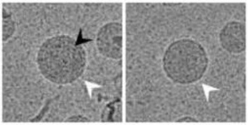
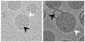
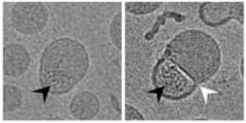
Each particle is assigned to one of three classes, defined by internal density and compartment structure, and reported as population fractions [4]:
Solid Core particles usually represent the major population of typical LNP preparations. The particles exhibit a near spherical appearance and an overall homogeneous, solid core central density. The outermost lipid layer(s) (white arrow) are typically visible. Inner density fluctuations due to an accumulation of electron dense mass (i.e. nucleic acids) are sometimes visible (black arrow).
Biphasic Split or “split” particles exhibit two or more clearly separated compartments in one LNP. Each compartment is typically not separated by a prominent electron dense layer and may consist of solid core LNP material (white arrow) and very low density material (i.e. buffer) (black arrow), respectively. The Split in the particle may be small (left image), hence, hardly modifying the spherical appearance of the LNP, or more pronounced (right image), hence, introducing large bulges into the particle.
Biphasic Dense or “blebbed” particles exhibit two or more clearly separated, densely filled compartments in one LNP. Individual compartments are often enclosed by a clearly discernible electron dense layer (white arrow). Current scientific literature suggests that the mottled density (black arrow) represents accumulated nucleic acids, while the second phase consists of morphologically more homogeneous solid core LNP material.
Roundness, defined as 4π·area / perimeter², where 100% is a perfectly circular cross-section. Cryo-EM resolves this per particle and reports it as a full distribution; ensemble sizing methods do not measure it [4].
According to our internal data, smaller, rounder particles with a higher Solid-Core fraction are the better-performing population [4].
In our “The Optimal Particle” field guide, we back this claim with data, one structural relationship at a time.
Cryo-EM can provide structural insights that other methodologies simply cannot resolve, providing critical data to inform development decisions the team needs to make, including:
ATEM works with biotech and pharmaceutical partners through GMP and non-GMP cryo-electron microscopy workflows, collaborating closely with scientific teams throughout research, preclinical, and clinical development. And our cryo-EM result doesn’t end with a morphology report. At ATEM, we interpret the structural differences according to the development decision the R&D team needs to make.
If your next LNP characterization project is already defined and you are looking for a data analysis partner, you can talk directly with our scientists about our cryo-EM services.
[1] Chen, S., et al. (2016). Influence of particle size on the in vivo potency of lipid nanoparticle formulations of siRNA. Journal of Controlled Release, 235: 236-244. doi:10.1016/j.jconrel.2016.05.059
[2] Hassett, K.J. et al. (2021). Impact of lipid nanoparticle size on mRNA vaccine immunogenicity. Journal of Controlled Release, 335: 237–246. doi:10.1016/j.jconrel.2021.05.021
[3] Crawford, R., et al. (2011). Analysis of lipid nanoparticles by Cryo-EM for characterizing siRNA delivery vehicles. Int J Pharm, 403(1-2): 237-244.
[4] ATEM (2026). The optimal particle field guide.
You are currently viewing a placeholder content from Vimeo. To access the actual content, click the button below. Please note that doing so will share data with third-party providers.
More InformationYou are currently viewing a placeholder content from YouTube. To access the actual content, click the button below. Please note that doing so will share data with third-party providers.
More InformationYou need to load content from reCAPTCHA to submit the form. Please note that doing so will share data with third-party providers.
More Information