Explore our cryo-EM applications for drug discovery & analytics
Cryo-EM: One method — a myriad of solutions
We offer a wide range of solutions for applications in the pharmaceutical and biotech industry. Have a look at how our cryo-EM technology can help aid your drug discovery or development projects.
Discovery
3D Structure determination: From protein & ligand to 3D Structure in a single fully integrated commercial facility.
Extensive Experience: Leverage over 50 years of combined cryo-EM expertise.
Comprehensive capabilities: Take advantage of our broad range of biochemical services to increase the success of your structure determination project.
Analytics
GMP facility: Offering the analysis of your samples in our GMP certified facility.
Proprietary Software: We transform simple (cryo-) EM image data with our (AI) Software into fully quantitative insights.
Streamlined processes: With an integrated platform & software we offer rapid turnaround times for your samples.
Services
Unlock the fundamentals of your protein. We tackle targets that are difficult to crystalize – including large assemblies or membrane proteins.
Lipid-Nano-Particle Characterization
Our AI-powered solutions offer unprecedented quantitative data into the morphology of lipid nanoparticles. Cryo-EM offers observable results, something impossible with indirect methods like Dynamic Light Scattering (DLS). This combination enables robust formulation and forced degradation studies.
Benefit from our unique, high-throughput cryo-EM platform, and discern challenging epitopes in 3D. With the possibilities of cryo-EM, you can screen several FAB/mAbs in complex with your target to visualize conformations, contacting residues, and binding locations.
We provide control over the cryo-EM 3D structure determination process. With our cutting-edge cryo-EM facility, computational resources, and 50+ years of collective expertise, we optimize and de-risk project budget. Our unique biochemical sample optimization assays enhance project success rates.
Cryo-EM revolutionizes AAV characterization in gene therapy R&D, offering precise analysis of critical quality attributes. It enables unparalleled identification of full, empty, and intermediate AAV particles across various serotypes, enhancing quality assurance and therapeutic efficacy.
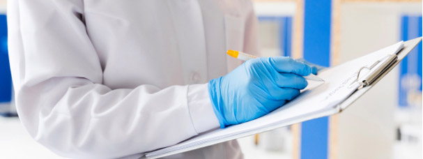
As a high-tech cryo-electron microscopy (cryo-EM) service provider, we possess over 50 years of expertise, and are GMP certified. Our unique selling point is our proprietary AI solutions, complementing a comprehensive biochemistry infrastructure, cutting-edge cryo-EM hardware, and a high-performance computing cluster. Our services are dedicated exclusively to biotech and pharmaceutical companies, supporting their research and development efforts. Combining advanced technology and in-depth knowledge, we empower innovation in these crucial sectors.
In-depth capabilities & expertise
For the characterization of bilogics we offer robust and high quality sample preparation procedures in our GMP certified lab. Ensuring highest industrial standards at unrivaled efficiency.
High-resolution 3D Structure determination requires highes quality proteins to succeed. In case client samples do not immidietly meet those requirements we have years of experience, broad cryo-EM specialized toolset to successfully ensure cryo-readiness for customer proteins.
Our fully integrated laboratory houses dedicated R&D and GMP wetlab facilities offer everything, among others chromatographic, ultracentrifugation and walkin-fridge capacities for optimal sample handling.
Industry dedicated propriatary AI software
Our core strenght is the integration of top sample preparation and fully controlled data acquisiton integrated in own industry purpose developed AI software tools.
We cover everything from GMP analytical software for Cell Line Characterization, AAV fillrate anaylsis to automated 3D structure determination workflows.
The science behind single-particle Cryo-EM
See all the steps of single-particle cryo-EM from protein production to finalized structure.
01
Protein Purification
02
Cryo-grid Preparation
03
Cryo-grid Freezing
04
Data Collection
05
Analysis and Processing
06
Structure Determination
Feasibility Study
Your cryo-EM structure starts with sample preparation. Once we receive your purified sample (1), we quickly process it with a negative stain analysis. This allows us to check sample quality and get back to you with a report on how the samples look in the microscope.
Cryo-EM
After the sample’s quality control, we proceed to prepare the grids (2). We do this by adding a small amount (only a few µl) of your sample to a cryo-grid and blotting it to achieve a thin layer of liquid. The grid is then vitrified in liquid ethane (3) so rapidly that the sample retains its native state without forming ice crystals.
Processing
Our specialists process the data quality and start collecting images (4). These images are analyzed and processed according to our standardized methods (5). We start 3D reconstruction and create a final structure from the density map (6). In the end, you receive an extensive report with the 3D structure as a PDB file.
The Rise of Cryo-EM
High-resolution cryo-EM structures
Ligand binding
Epitope mapping
Lipid nanoparticles
Plasmid DNA
Viral particle analysis
Viral vectors
Join the resolution revolution
Since the 1950s, scientists laid the foundations for visualizing protein structures. They gazed upon the unseen and unlocked the hidden nature of protein biology by combining innovative machinery together with human ingenuity. The first protein structure was obtained using x-ray crystallography, a method that relies on sending x-ray beams through proteins in a crystalized lattice. This method has dominated the structural biology field until recently.
Cryogenic electron microscopy (cryo-EM) uses beams of electrons to highlight proteins in a variety of orientations. One can think of it like a projector that shines on an object, creating contrast in the light and showing the object’s shape. With thousands or millions of different orientations, scientists can combine and reconstruct the shapes in a single, 3-dimensional view. While being a substantial method, cryo-EM fought to provide the same high-resolution structures of x-ray crystallography due to hardware limitations. In recent years, this has changed, with advances in cryo-EM leading to the so-called “resolution revolution”.
The resolution revolution shaped cryo-EM into a powerful method that now competes closely with x-ray crystallography when it comes to resolution. It provides certain benefits over x-ray crystallography by requiring much lower sample amounts and targeting challenging projects such as complexes and membrane proteins, with higher reliability. Since protein samples are vitrified instead of crystallized, it allows a look into how the protein behaves in solution. In other words—we can see the natural dynamics that govern protein function.
One method – multiple aspects
Cryo-EM takes on challenges with ease. Membrane proteins, large complexes, and flexible proteins—all are applicable candidates. With the natural, soluble state of the protein sample, it is possible to visualize and determine the binding of ligands and antibodies. Proteins are often flexible by nature, transporting chemicals into cells or responding to cascades that trigger complicated events. With cryo-EM, these flexible traits can be determined. Important mutations can be discerned and compared to their wild-type counterpart, providing atomic details of how diseases work. Antibodies can be developed with high certainty by locating their binding domains and epitopes on the antigen.
Cryo-EM and transmission electron microscopy (TEM) are not only used to discern structural data. Analytics combines visual and quantitative data by analyzing lipid nanoparticles (LNP) and adeno-associated virus (AAV) delivery systems. We test cellular samples with TEM for viral particle analysis, ensuring certainty and safety before drug release testing. TEM offers a unique addition to other methods by visualizing samples and determining the quality by ruling out contamination and unfavorable morphology – all in one single analysis.
We at ATEM focus entirely on electron microscopy. Our scientific team consists of specialists that together combine decades of work in protein biochemistry, cryo-EM, and data engineering. Our passion is reflected in our results, and we dedicate ourselves to reliability and transparency in order to take on the challenges of drug design and scientific discovery.
Cryo-EM
Sample quantity: low / medium
Protein / Complex size: small - very large
Native state: yes
Dynamics visualization: yes
Need for crystallization: no
X-Ray Crystallography
Sample quantity: high
Protein / Complex size: high
Native state: no
Dynamics visualization: no
Need for crystallization: no
NMR
Sample quantity: high
Protein / Complex size: small
Native state: yes
Dynamics visualization: yes
Need for crystallization: yes
Cryo-EM
X-Ray Crystallography
NMR
Sample amount
low / medium
high
high
Protein/Complex size
>50 kDa
small-large
<50 kDa
Native state
yes
no
yes
Protein dynamics
yes
no
yes
Need for crystallization
no
yes
no
You need to load content from reCAPTCHA to submit the form. Please note that doing so will share data with third-party providers.